

DETECTION OF FOCAL LIVER DISEASES FROM ULTRASOUND IMAGES
Computerized image analysis and recognition for malignant liver tumors detection
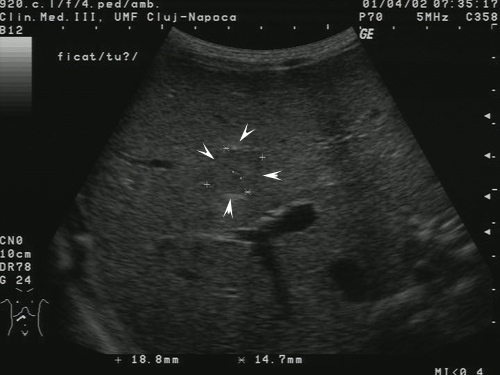
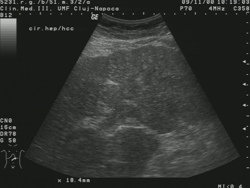
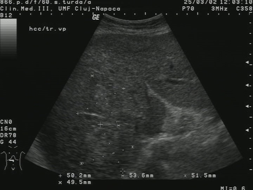
Liver chronic diseases constitute an important public health issue. The evolution of diffuse liver diseases is variable, but it has generally long term. Whatever the nature of the liver aggression, it seems to follow a pattern characterised by the successive stages: inflammation (at the beginning), necrosis, fibrosis, regeneration (cirrhosis), dysplasia, and hepatocellular carcinoma . At the end, the Hepatocellular carcinoma (HCC) is one of the most frequent malignant tumors of liver (75% of the liver cancer cases). Other well known malignant liver tumors are hepatoblastoma (7%), cholangiocarcinoma and cystadenocarcinoma (6%) [7]. Eloquent images of HCC are illustrated bellow:
|
Incipient HCC |
|
||
|
Mature HCC – |
|
|
|
|
Encephaloid form |
Diffuse form |
Multicentric form |
|
Objectives
As the classical method – the liver and tumors biopsy, can be very dangerous for the patients, we aim to develop non-invasive, computerized methods for image analysis and recognition. Texture-based image analysis is considered very appropriate for this purpose. Thus, the goals are:
• Build the imagistic textural model of HCC consisting in:
• Automatic recognition of HCC, especially in incipient phases or even forerunner phases [9]• The relevant textural features, correlated with the visual characteristics of hepatocellular carcinoma in its various evolution stages, that make the difference between: normal liver and hepatocellular carcinoma; cirrhosis and hepatocellular carcinoma [4], [5], [6], [10]
• The characteristic values of the textural features in the case of HCC
Methodology
Image acquisition: Images
are acquired through a General Electric, Logiq 7 ecograph using a well
established protocol concerning the device settings. All the images
from a certain training set are obtained using the same settings of the
echographic device in order to eliminate irrelevant, confusing
differences between classes.
Image analysis: The
image analysis consists in two phases – the computation of the textural
features on the region of interest, then the establishment of the
exhaustive set of relevant textural features. The computation of
textural features is done through specific texture analysis methods:
- • the first order statistics of grey levels [1], [2]
- • the Grey Level Co-occurrence Matrix (GLCM) as a second order statistic and its associated parameters: correlation, contrast, energy, entropy, local homogeneity [2]
- • Fractal-based methods: Box-Counting method, Hurst algorithm [3]
- • Transform-based methods: Wavelet Transform, Gabor Transform [8], [11]
In order to obtain the exhaustive set of relevant textural features the following methods are taken into consideration:
-
- • univariate density modeling - in order to detect the bimodal features, that could influence the class parameter [6]
- • classifiers that include the extraction of relevant features - (Bayesian Belief Networks, Decision Trees , AdaBoost , Support Vector Machine) [4], [5], [10]
- • methods for the analysis of the mutual influence that exist between the features (regression) [4], [6]
- Automatic recognition: The possibility of automatic (fully computerized) recognition of HCC, using the relevant features, is also studied. The following classification methods are considered for this purpose: Bayesian classifiers, Artificial Neural Networks (ANN), the Multilayer Perceptron method, Decision Trees, AdaBoost, Support Vector Machines (SVM) [4], [5], [10].
Achievements
• Textural features computation, plotting and comparison• The imagistic textural model of HCC
• The exhaustive set of independent relevant textural features for HCC characterization :
• The specific values of the relevant textural features in the case of HCC:• GLCM entropy, GLCM homogeneity, average value of grey levels, edge frequency, edge contrast, the autocorrelation index, the Hurst coefficient, the entropies computed after applying the Wavelet Transform - characterization of the complexity in the grey levels structure
• High accuracy of HCC automatic recognition: about 90%• The average of the grey levels takes low values
• GLCM entropy, GLCM homogeneity, Edge contrast, edge frequency, the autocorrelation index, the Hurst coefficient and the entropies computed after applying the Wavelet Transform take high values
Illustrations
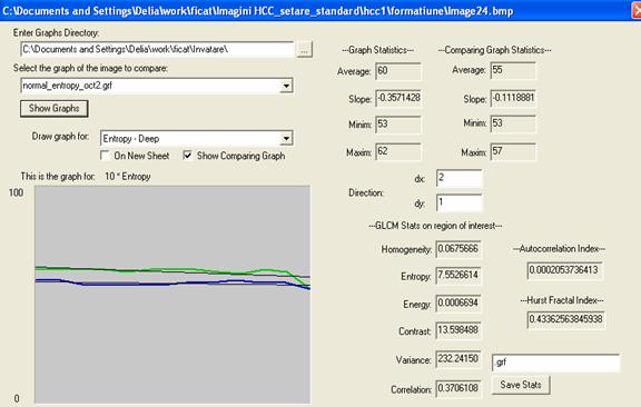
Fig.2. Textural features computation, plotting and comparison: the GLCM entropy is higher in the case of HCC (green color) than in the normal case (blue color)
|
maxg <= 74 | | | Hurst <= 0.095744: no (6.0)
| | | Hurst > 0.095744: yes (5.0/1.0) |
Decision Tree : the relevance of textural features in HCC characterization |
|
|
|
Bayesian Belief Network: independent textural parameters that influence the HCC diagnosis |
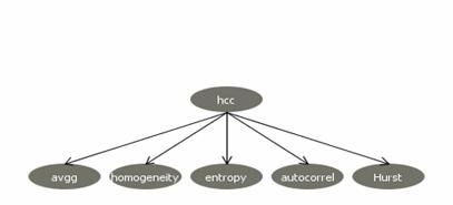
Fig.3. Textural parameters analysis for relevant features identification
Future goals
- Improving the experiments by collecting a larger number of
cases/class and making the comparison also with the other malignant
tumors and the benign liver tumors
- Automatic segmentation of HCC using the set of relevant textural parameters
References
[1] Clausi, D.A. (2002), “An analysis of co-occurrence texture statistics as a function of grey level quantization”, Canadian Journal of Remote Sensing, vol. 28, no. 1, 2002, pp. 45-62
[2] Chen, S., K. Cheng, Y. Dai,Y.Sun, Y. Chen, Y. Chang, “The representation of sonographic image texture for breast cancer using co-occurrence matrix”, Journal of Medical and Biological Engineering , vol. 25, no.4, 2005, pp.193-199
[3] Chikui, T., K. Tokumori, K. Yoshiura, K. Oobu, S.Nakamura, K. Nakamura, “Sonographic characterization of salivary gland tumors by fractal analysis” , Ultrasound in Medicine and Biology, vol. 31, no. 10, 2005, pp. 1297–1304
[4]Duda, R., P. E. Hart and D. G. Stork (2000); Pattern Classification (2nd ed), Wiley Inter-science, 2003
[5] Hall, M., G. Holmes, “Benchmarking Attribute Selection Techniques for Discrete Class Data Mining”, IEEE Transactions on Knowledge and Data Engineering, vol. 15, no.3, 2003, pp. 1 – 16
[6] Jain A K, R. Duin, J. Mao, “Statistical Pattern Recognition: A Review”, IEEE Transactions on Pattern Analysis and Machine Intelligence, vol. 22, no.1, January 2000, pp. 4-37
[7] Sherman, M., MB, BCh, PhD, “Approaches to the Diagnosis of Hepatocellular Carcinoma”, Current Gastroenterology, Reports 2005, Current Science Inc.
[8] Stollnitz, E., Tony D. DeRose, and David H. Salesin, “Wavelets for computer graphics”,. IEEE Computer Graphics and Applications, vol. 15, no. 3, May 1995, pp.76–84
[9] Sujana, H., S. Swarnamani and S. Suresh, “Application of
Artificial Neural Networks for the classification of liver lesions by
texture parameters”, Ultrasound in Med.[[[[&]]]] Biol., vol. 22, no. 9, 1996, pp. I177- 1181,
[10] Witten, I., E. Frank, “Data Mining: Practical machine learning tools and techniques,” 2nd Edition, Morgan Kaufmann, San Francisco, 2005
Publications
D. Mitrea, S. Nedevschi, C. Cenan, M. Lupsor, R. Badea, "Exploring Texture-Based Parameters, Noninvasive Characterization and Modelling of Diffuse Liver Diseases and Liver Cancer from Ultrasound Images", WSEAS Transactions on Computers, vol. 6, no.2, February 2007, pp. 283-291
D. Mitrea, S. Nedevschi, M. Lupsor, R. Badea, "Texture-based approach for Building the Imagstic Model of Hepatocellular Carcinoma", Proceedings of The 13th International Symposium on System Theory, Automation, Robotics, Computers, Informatics, Electronics and Instrumentation (SINTES13), Craiova, Romania, October 18-20, 2007, pp.68-73
M. Lupsor, D. Mitrea, S. Nedevschi, R. Badea, "Representation and modeling of cirrhosis and hepatocellular carcinoma from ultrasound images using texture-based methods", presented at the European Congress of Echography, EUROSON, Leipzig, Germany, October 24-27, 2007; published in Ultraschall in der Medizin - European Journal of Ultrasound, 2007, vol. 28, pp. S4
H. Stefanescu, R. Badea, M. Lupsor, S. Tripon, O. Dancea, D. Capatana, I. Stoian, D. Mitrea, T. Marita, S. Nedevschi, L. Neamtiu, V. Popita., "Telemedicine network for ultrasound screening of HCC", Ultraschall in der Medizin - European Journal of Ultrasound, 2007, vol. 28, pp. S59
D. Mitrea, S. Nedevschi, P. Mitrea, M. Lupsor, R. Badea, I. Coman, "Exploring texture-based parameters for the automatic recognition of the hepatocellular carcinoma (HCC) and prostatic adenocarcinoma (ADKP) from ultrasound images", poster presented at the CEEX Conference, Sibiu, Romania, October 25-26, 2007
R. Badea, H. Stefanescu, M. Lupsor, Z. Sparchez, H. Branda, T. Pop, S. Tripon, M. Grigorescu, S. Nedevschi, I. Stoian, V. Popita, D. Mitrea, D. Capatana, O. Dancea, O. Suteu, L. Neamtiu, "Screening and Surveillance in Liver Cirrhosis. Actual Trends for the Early Detection of Hepatocellular Carcinoma. Is TELEHEPASCAN Project a Viable Option?" Romanian Journal of Hepatology vol. 2, no.1, 2006, pp. 27-35.
D. Mitrea, S. Nedevschi, M. Lupsor, R. Badea, "Texture-Based Methods for Noninvasive Evaluation of Diffuse Liver Diseases, Liver Cancer and Prostate Cancer from Ultrasound Images", The 6-th Joint Conference on Mathematics and Computer Science (MACS '06), Pecs, Hungary, July 12-15, 2006, pp. 65